Artroskopija kuka
dr. sc. Tomislav Čengić, ortoped.png)
T. Čengić, D. Jurković, H. Hajsok, T. Smoljanović, L. Novosel, K. Rotim i D. Delimar
Artroskopija kuka je minimalno invazivni, učinkovit i inovativni ortopedski zahvat s niskim rizikom od komplikacija. Kod našeg bolesnika ostatna cam lezija i fragment kosti koji je zaostao u prednjem dijelu čahure kuka nakon artroskopije kuka izvedene tri godine ranije izazvao je utrnulost natkoljenice, fascikulacije mišića i parestezije. Pretpostavljamo da je koštani ulomak zaostao kao komplikacija brušenja kosti u prethodnom zahvatu. Prilikom fleksije kuka komprimirao je živčane strukture i posljedično izazivao spomenute simptome. Komplikacija je riješena revizijskom artroskopijom kuka i uklanjanjem slobodnog fragmenta. Nakon revizijske operacije došlo je do smanjenja boli i prestanka utrnulosti prednjeg dijela bedra u našeg bolesnika.
Ključne riječi: Artroskopija kuka; Artroskopija, Lezija cam; Slobodni fragment; Utrnulost bedra; Fascikulacije mišića; Parestezije
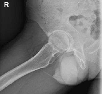
Fig. 1. Preoperative radiographs of the right hip with residual anterolateral cam impingement (black arrow) and a small bone fragment (white arrow); lateral frog view.
.png)
Fig. 2. Preoperative pelvic multi-slice computerized tomography with residual cam deformity (white arrow) of the right hip and a small bone fragment (orange arrow); (a) frontal plane view; (b) horizontal plane view; (c) three-dimensional reconstruction (with capsular loose fragment depicted by white arrow).
.jpg)
.jpg)
.jpg)
.jpg)
Fig. 3. Hip arthroscopy procedure intraoperative photos: (a) residual cam deformity before debridement; (b) debrided cam deformity; (c) intracapsular loose bony fragment before mobilization; (d) mobilized loose bony fragment before extraction.
HIP ARTHROSCOPY: RESIDUAL CAM DEFORMITY COMBINED WITH LOOSE BONY FRAGMENT IN HIP CAPSULE
Running title: Hip arthroscopy – cam lesion
Tomislav Čengić1,2, Danijel Jurković1, Hana Hajsok1, Tomislav Smoljanović3,4, Luka Novosel5, Krešimir Rotim2,4,6 and Domagoj Delimar3,4
1Department of Traumatology, Sestre milosrdnice University Hospital Center, Zagreb, Croatia;
2University of Applied Health Sciences, Zagreb, Croatia;
3Department of Orthopedic Surgery, Zagreb University Hospital Center, Zagreb, Croatia;
4School of Medicine, University of Zagreb, Zagreb, Croatia;
5Department of Radiology, Sestre milosrdnice University Hospital Center, Zagreb, Croatia;
6Department of Neurosurgery, Sestre milosrdnice University Hospital Center, Zagreb, Croatia
Correspondence to: Tomislav Čengić, MD, PhD, Department of Traumatology, Sestre milosrdnice University Hospital Center, Draškovićeva 19, HR-10000 Zagreb, Croatia
E-mail: cengict@me.com
Received October 27, 2021, accepted December 11, 2021
Introduction
Hip arthroscopy is used as a diagnostic and therapeutic method in the treatment of various intra-articular and extraarticular hip conditions. As the procedure is technically quite demanding, it is connected with a higher complication rate compared to arthroscopy of other human joints1,2. The most common causes for hip arthroscopy revision are labral tears and femoroacetabular impingement (FAI)3. There was no presentation of hip arthroscopy revision due to loose bony fragment in the anterior hip capsule.
A revision hip arthroscopy due to residual cam lesion combined with loose bony fragment in the anterior hip capsule causing limited range of motion, pain, muscle fasciculations and numbness in the thigh is reported.
Case Report
A 22-year-old man with a history of right-side hip pain was examined in February 2021. The patient underwent arthroscopic resection of cam lesion of the right hip at another institution three years before. Due to increased pain in the operated hip, multiple courses of physical therapy and non-steroidal anti-inflammatory drugs (NSAIDs) were prescribed to the patient. As the effort failed to reduce the symptoms, he became anxious and depressed. His primary care provider prescribed him antidepressants and referred him to our department.
An analgesic gait was present on his arrival. The pain was getting worse after long sitting. There was no history of trauma following the surgery. On clinical examination, paresthesia over anterior thigh and fasciculation of quadriceps muscle were revealed on deep palpation over anterior capsule of the affected hip. Flexion of the hip was limited to 90° and internal rotation to 10° due to strong pain. Initial plain radiographs, anteroposterior and lateral frog view suggested residual anterolateral cam type of impingement and small bone fragment (Fig. 1). Computed tomography (CT) scan demonstrated findings of a sclerotic zone in the lateral part of the right femur head and a small bone fragment was noticed in the anterior part of the hip joint on three-dimensional reconstruction scan of the pelvis (Fig. 2). Due to suspected osteoid osteoma (OO), a tumor of the femoral neck, the patient was first referred to invasive radiologist for CT guided biopsy analysis of the bone, followed by radiofrequency ablation therapy (RFA). Biopsy results excluded the presence of the OO.
Following the RFA procedure, the pain decreased for a few days, probably due to local anesthetic that was used, and thereafter the pain steadily increased. Because of the limited range of movement and residual strong pain in the anterior part of the right hip, the patient opted for revision hip arthroscopy.
The procedure was performed in October 2021 in supine position, according to the procedure described by Thaunat et al.4. The anterior and anterolateral portals with standard length arthroscope were used, without traction of the hip. After debridement of abundant fibrosis tissue in the hip joint, a small loose bone fragment in the anterior hip capsule was noticed. While using electrocautery for the loose fragment debridement, fasciculations of the quadriceps muscle occurred. Electrocautery debridement was immediately converted to debridement without electrostimulation using arthroscopic hook and shaver. Bone fragment was separated from the anterior capsule and extracted from the joint. A cam lesion was identified in the anterosuperior and posterolateral region of the head. The prominent head-neck cam lesion was removed with the arthroscopic burr (Fig. 3).
Considerable resolution of pain and loss of anterior thigh numbness on palpation was reported on the first day after the revision surgery. The patient was discharged on the second postoperative day with recommendation to take indomethacin 25 mg orally twice a day for 3 weeks and low-molecular-weight heparin (LMWH) once daily subcutaneously for 4 weeks. Moreover, the patient was advised protection from weight bearing for 4 weeks. On the first follow up, two weeks after the surgery, during hip flexion to 90°, numbness and fasciculations of the quadriceps muscle were completely absent.
At the two-month follow up, the pain subsided, as rated on the visual analog scale (VAS 2). On clinical examination, the FABER test was positive. On the other hand, there were no fasciculations of the quadriceps muscle, FADIR test was negative, and palpation of the thigh was painless.
Discussion
A bone fragment remaining in the front part of a hip after hip arthroscopy caused thigh numbness and paresthesia. The complication was resolved by revision hip arthroscopy which, in addition to cam bump resection, removed the bone fragment from the capsule. In our case report, we assume that the loose bony fragment was a complication of burring on the previous procedure. During hip flexion, femoral nerve was compressed and consequently caused thigh paresthesia. One of the options we considered as a cause was heterotopic ossification (HO). In their meta-analysis of 6334 hips from 92 studies, Harris et al. report that the complication rate after hip arthroscopy was 7.5% of minor complications and 0.58% of major complications. The most common complication was associated with iatrogenic chondrolabral injury. More than 400 reoperations were performed after hip arthroscopy, of which 30% were arthroscopic. The most common cause for arthroscopic reoperation was loose body removal and lysis of adhesions3. Sampson found 6.4% complication rate in 530 cases among several experienced surgeons and those reported in literature, yet none of the complications described fits our case5. Kowalczuk et al. report a complication rate of 4.0%, having reviewed 66 studies. Notably, in more than 10 cases, there was a bony fragment causing FAI6,7.
Nakano et al. in their meta-analysis of 36761 hips report that 1221 complications occurred during or after hip arthroscopy, yielding an overall complication rate of 3.3% with major complication rate of 0.16%. The most commonly reported complication was neurapraxia, followed by iatrogenic chondrolabral injury. In addition, 2.9% of complications were due to loose or foreign fragments8.
Randelli et al. reviewed 300 cases of femoroacetabular impingement treated by hip arthroscopy and found five cases of HO (1.6%)9. Byrd and Jones found HO of the articular capsule in one of 207 patients having undergone arthroscopic procedure for femoroacetabular impingement10. Prophylaxis against HO in patients who underwent hip arthroscopy can come in a variety of forms; however, the most common is NSAID therapy. RFA can also be used. Considering that, our patient was administered NSAIDs. Beckman et al., prospectively evaluated the effect of NSAIDs in preventing HO after hip arthroscopy and found that the incidence of HO in cases in which patient did not receive NSAID prophylaxis was 25% (23/92), compared to 5.6% (11/196) of cases in which patient received NSAIDs. The authors conclude that routine NSAID prevention reduces but does not limit the incidence of HO in patients undergoing hip arthroscopy11. As known from medical history, our patient did not receive NSAID prophylaxis after the primary hip arthroscopy procedure.
Nerve injury during hip arthroscopy is a well-documented complication. Most of them are minor complications. Previous literature suggests a wide range of incidence from 0.5% to 5%12. In their prospective study, Kern et al. report that 13% of 100 patients had a nerve injury. Specific nerve injury included the pudendal, lateral femoral cutaneous, sciatic and superficial peroneal nerve8,13-15. All of the cases resolved spontaneously. We found no case in the literature with 3-year postoperative numbness. Nerve injuries are mostly associated with traction time during the procedure. Telleria et al. studied the prevalence of sciatic nerve injury during hip arthroscopy using intraoperative nerve monitoring in 60 patients. They found intraoperative nerve deficit in 58% of patients and postoperative clinically evident sciatic nerve injury in 7% of patients. The average traction time of 32 minutes was responsible for the onset of sciatic nerve injury16. Nevertheless, when the portals are placed more medially, there is a risk for the lateral femoral cutaneous nerve and its branches to be injured17,18. At a venture, only one femoral palsy has been reported in the literature19.
Several studies have shown that FAI (in 74.8%) and insufficient resection are the main reasons for revision arthroscopy of the hip17,20. The patient presented in this report had residual cam deformity that might contributed to the limited range of motion and pain on hip flexion. However, we are not aware that cam deformity can cause paresthesia and numbness of the thigh, as in our case. Being in the hip for the second time, the remaining cam deformity was removed, and one could speculate that resection decreased paresthesia and numbness as well.
Limitations of this study include the lack of electromyoneurography confirmation of the femoral nerve irritation and lack of follow up magnetic resonance imaging scan that would visualize the relationship between the bone fragment and femoral nerve. However, considering the patient’s symptoms prior to revision hip arthroscopy, intraoperative quadriceps muscle fasciculations occurring when electrocautery was used for the loose fragment debridement and loss of neurological symptoms following revision hip arthroscopy, we are convinced that the femoral nerve irritation was the cause of problems in the patient.
Conclusion
When performing hip arthroscopy, a surgeon has to be careful to remove bone fragments completely from the patient’s body, i.e., not to leave them within or outside the joint capsule, as they can irritate the nearby nerve. In case of paresthesia in the front part of a thigh following hip arthroscopy, one should seek for the remaining bone fragment that might cause nerve irritation. Revision hip arthroscopy can provide a solution for patients with symptomatic residual cam deformity and/or unexplainable paresthesia of the thigh.
References
1.Mehta N, Chamberlin P, Marx RG, et al. Defining the learning curve for hip arthroscopy: a threshold analysis of the volume-outcomes relationship. Am J Sports Med. 2018;46(6):1284-93. doi: 10.1177/0363546517749219
2.Minkara AA, Westermann RW, Rosneck J, Lynch TS. Systematic review and meta-analysis of outcomes after hip arthroscopy in femoroacetabular impingement. Am J Sports Med. 2019;47(2):488-500. doi: 10.1177/0363546517749475
3.Harris JD, McCormick FM, Abrams GD, et al. Complications and reoperations during and after hip arthroscopy: a systematic review of 92 studies and more than 6,000 patients. Arthroscopy. 2013;29(3):589-95. doi: 10.1016/J.ARTHRO.2012.11.003
4.Thaunat M, Murphy CG, Chatellard R, et al. Capsulotomy first: a novel concept for hip arthroscopy. Arthrosc Tech. 2014;3(5):e599-e603. doi: 10.1016/J.EATS.2014.06.016
5.Sampson TG. Complications of hip arthroscopy. Clin Sports Med. 2001;20(4):831-6. doi: 10.1016/S0278-5919(05)70288-X
6.Badylak JS, Keene JS. Do iatrogenic punctures of the labrum affect the clinical results of hip arthroscopy? Arthroscopy. 2011;27(6):761-7. doi: 10.1016/J.ARTHRO.2011.01.019
7.Kowalczuk M, Bhandari M, Farrokhyar F, et al. Complications following hip arthroscopy: a systematic review and meta-analysis. Knee Surg Sports Traumatol Arthrosc. 2013;21(7):1669-75. doi: 10.1007/S00167-012-2184-2
8.Nakano N, Lisenda L, Khanduja V, Jones TL, Loveday DT. Complications following arthroscopic surgery of the hip: a systematic review of 36 761 cases. Bone Joint J. 2017;99-B(12):1577-83. doi: 10.1302/0301-620X.99B12.BJJ-2017-0043.R2
9.Randelli F, Pierannunzii L, Banci L, Ragone V, Aliprandi A, Buly R. Heterotopic ossifications after arthroscopic management of femoroacetabular impingement: the role of NSAID prophylaxis. J Orthoph Traumatol. 2010;11(4):245-50. doi: 10.1007/S10195-010-0121-Z
10.Byrd JWT, Jones KS. Arthroscopic management of femoroacetabular impingement in athletes. Am J Sports Med. 2011;39:7S-13S. doi: 10.1177/0363546511404144
11.Beckmann JT, Wylie JD, Kapron AL, Hanson JA, Maak TG, Aoki SK. The effect of NSAID prophylaxis and operative variables on heterotopic ossification after hip arthroscopy. Am J Sports Med. 2014;42(6):1359-64. doi: 10.1177/0363546514526361
12.Nepple JJ, Byrd JWT, Siebenrock KA, Prather H, Clohisy JC. Overview of treatment options, clinical results, and controversies in the management of femoroacetabular impingement. J Am Acad Orthop Surg. 2013;21:S53-S58. doi: 10.5435/JAAOS-21-07-S53
13.Sing DC, Feeley BT, Tay B, Vail TP, Zhang AL. Age-related trends in hip arthroscopy: a large cross-sectional analysis. Arthroscopy. 2015;31(12):2307-13.e2. doi: 10.1016/J.ARTHRO.2015.06.008
14.Kern MJ, Murray RS, Sherman TI, Postma WF. Incidence of nerve injury after hip arthroscopy. Am Acad Orthop Surg. 2018;26(21):773-8. doi: 10.5435/JAAOS-D-17-00230
15.Magrill ACL, Nakano N, Khanduja V. Historical review of arthroscopic surgery of the hip. Int Orthop. 2017;41(10):1983-94. doi: 10.1007/S00264-017-3454-X
16.Telleria JJ, Safran MR, Harris AH, Gardi JN, Glick JM. Risk of sciatic nerve traction injury during hip arthroscopy—is it the amount or duration? An intraoperative nerve monitoring study. J Bone Joint Surg Am. 2012;94(22):2025-32. doi: 10.2106/JBJS.K.01597.
17.Philippon MJ, Schenker ML, Briggs KK, Kuppersmith DA, Maxwell RB, Stubbs AJ. Revision hip arthroscopy. Am J Sports Med. 2007;35(11):1918-21. doi: 10.1177/0363546507305097
18.Griffin DR, Villar RN. Complications of arthroscopy of the hip. J Bone Joint Surg Br. 1999;81(4):604-6. doi: 10.1302/0301-620X.81B4.9102
19.Salas AP, O’Donnell JM. Prospective study of nerve injuries associated with hip arthroscopy in the lateral position using the modified portals. J Hip Preserv Surg. 2016;3(4):278-87. doi: 10.1093/JHPS/HNW032
20.Thomas Byrd JW. Complications associated with hip arthroscopy. Operative Hip Arthroscopy. Published online 2005:229-35. doi: 10.1007/0-387-27047-7_16
21.Griffin JW, Weber AE, Kuhns B, Lewis P, Nho SJ. Imaging in hip arthroscopy for femoroacetabular impingement: a comprehensive approach. Clin Sports Med. 2016;35(3):331-44. doi: 10.1016/J.CSM.2016.02.002